Major Blood Vessel Chart - Blood Vessels Circulatory Anatomy - Arteries serving the upper limbs as the subclavian artery exits the thorax into the axillary region, it is renamed the axillary artery.
Major Blood Vessel Chart - Blood Vessels Circulatory Anatomy - Arteries serving the upper limbs as the subclavian artery exits the thorax into the axillary region, it is renamed the axillary artery.. The catheter is then threaded through the major blood vessels and into the chambers of the heart. If you were to lay out all the blood vessels of the body in a line, they would stretch for nearly 60,000 miles. There are three distinct layers, or. Blood is circulated through the body by blood vessels via the cardiovascular system which is comprised of the heart and the circulatory system.arteries move blood from the heart first to smaller arterioles, then capillaries or sinusoids, venules, veins, and back to the heart. Bulky middle tunic contains smooth muscle and elastin 3.
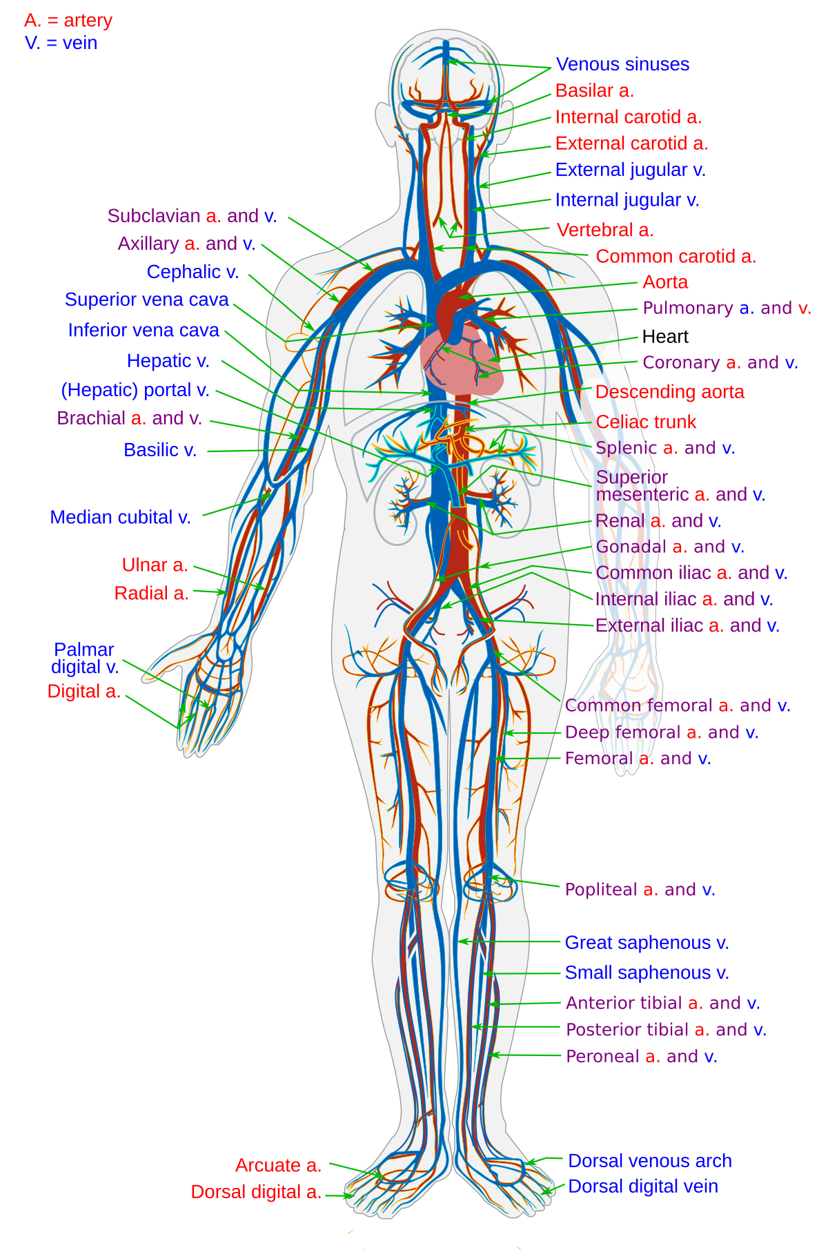
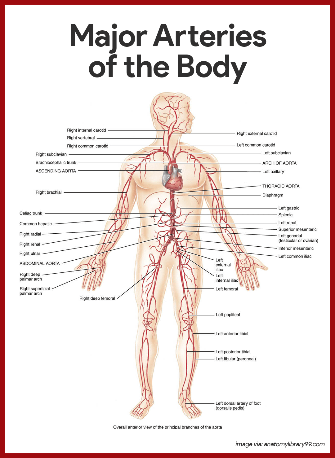
Use key choices to identify the blood vessel tunic described. Arteries (in red) are the blood vessels that deliver blood to the body. Blood flow through the heart. The major arteries in the body. The catheter is then threaded through the major blood vessels and into the chambers of the heart.
The major arteries in the body.
Artery, vein, and capillary •list the major disorders of blood vessels and explain how they develop •trace the path of blood through the systemic, pulmonary, portal, and fetal circulations. Beginning with the superior and inferior vena cavae and the coronary sinus, the flowchart below summarizes the flow of blood through the heart, including all arteries, veins, and valves that are passed along the way. Strokes can be isolated to the anterior or posterior circulation depending on the vessels affected. 175 body part artery vein heart aorta vena cava head carotid jugular vein arms subclavian artery subclavian vein kidney renal artery renal vein legs iliac artery iliac vein intestines mesenteric arteries hepatic vein pulmonary circuit right ventricle æ pulmonary artery æ lungs æ pulmonary veinæ left atrium. Fluid is also brought back to the heart via the lymphatic system. Superior and inferior vena cavae and the coronary sinus 2. Figures 1 and 2 show the major arteries and veins of the body. Its smooth surface decreases resistance to blood flow Create a flow chart showing the major systemic arteries through which blood travels from the aorta and its major branches, to the most significant arteries feeding into the right and left upper and lower limbs create a flow chart showing the major systemic veins through which blood travels from the feet to the right atrium of the heart The major arteries in the body. The catheter is then threaded through the major blood vessels and into the chambers of the heart. Arteries (in red) are the blood vessels that deliver blood to the body. The flow chart summarizes the distribution of the major branches of the common iliac arteries into the pelvis and lower limbs.
Vasomotor fibers • constriction of blood vessels raises blood pressure. The electrocardiogram (ekg or ecg) transport of co2 (carbon dioxide) in the blood Home » unlabelled » major blood vessel chart : Vessel elasticity blood volume cardiac output blood vessel diameter blood viscosity total vessel length peripheral resistance page 5. • vessel diameter is actively regulated by vasomotor fibers, sympathetic nerve fibers that innervate the vessel's smooth muscle layer.
Modified interlocking detachable coil indicated to obstruct or reduce rate of blood flow in the peripheral vasculature.
The major arteries in the body. Blood supply to the scalp. Create a flow chart showing the major systemic arteries through which blood travels from the aorta and its major branches, to the most significant arteries feeding into the right and left upper and lower limbs create a flow chart showing the major systemic veins through which blood travels from the feet to the right atrium of the heart Major blood vessels of the human body learn by taking a quiz; Eventually, the smallest arteries, vessels called arterioles, further branch into tiny capillaries, where nutrients and wastes are exchanged, and then combine with other vessels that exit capillaries to form venules, small blood vessels that carry blood to a vein, a larger blood vessel that returns blood to the heart. The flow chart summarizes the distribution of the major branches of the common iliac arteries into the pelvis and lower limbs. The walls of the arteries are tightly and closely bound to the. Arteries serving the upper limbs as the subclavian artery exits the thorax into the axillary region, it is renamed the axillary artery. • vessel diameter is actively regulated by vasomotor fibers, sympathetic nerve fibers that innervate the vessel's smooth muscle layer. Anatomy of blood vessels review sheet 32 261 microscopic structure of the blood vessels 1. The catheter is then threaded through the major blood vessels and into the chambers of the heart. There are three distinct layers, or. Venous blood flow chart internal jugular v coronary sinus subclavian v brachiocephalic v superior vena cava brachiocephalic v axillary v.
It ultimately becomes the subclavian vein at the lateral border of the first rib. The major (or great) blood vessels of the heart are the larger arteres and veins that attach to the atria and ventricles and transport blood to and from the systemic circulatory system and the pulmonary circulatory system. Online quiz to learn major blood vessels of the human body; The electrocardiogram (ekg or ecg) transport of co2 (carbon dioxide) in the blood Create a flow chart showing the major systemic arteries through which blood travels from the aorta and its major branches, to the most significant arteries feeding into the right and left upper and lower limbs create a flow chart showing the major systemic veins through which blood travels from the feet to the right atrium of the heart
Online quiz to learn major blood vessels of the human body;
Blood flow through the heart. Eventually, the smallest arteries, vessels called arterioles, further branch into tiny capillaries, where nutrients and wastes are exchanged, and then combine with other vessels that exit capillaries to form venules, small blood vessels that carry blood to a vein, a larger blood vessel that returns blood to the heart. Fluid is also brought back to the heart via the lymphatic system. The walls of the arteries are tightly and closely bound to the. The posterior auricular, occipital and superficial temporal arteries (along with two branches of the internal carotid artery; Deep veins, located in the center of the leg near the leg bones, are enclosed by muscle. Vessel elasticity blood volume cardiac output blood vessel diameter blood viscosity total vessel length peripheral resistance page 5. The catheter is then threaded through the major blood vessels and into the chambers of the heart. Arteries are a type of blood vessel. The decrease in blood flow can result from either obstruction of the blood vessels (atherosclerotic plaque formation) or rupture of a blood vessel (hemorrhagic stroke). It begins at the lower margin of the teres major muscle formed from the basilic vein and later the cephalic vein, gathering tributaries within the shoulders. Bulky middle tunic contains smooth muscle and elastin 3. Home » unlabelled » major blood vessel chart :


Komentar
Posting Komentar